
Dr. Scott Bradfield is a clinical trial principal investigator (PI), practicing oncologist, and Medical Advisor to Yunu. His unique perspective offers valuable insight into the day-to-day realities of clinical research, patient care, and the evolving role of technology in advancing clinical trials. As both a physician and researcher, Dr. Bradfield brings firsthand knowledge of the challenges and opportunities shaping the future of clinical development.
Dr. Scott Bradfield is a clinical trial principal investigator (PI), practicing oncologist, and Medical Advisor to Yunu. His unique perspective offers valuable insight into the day-to-day realities of clinical research, patient care, and the evolving role of technology in advancing clinical trials. As both a physician and researcher, Dr. Bradfield brings firsthand knowledge of the challenges and opportunities shaping the future of clinical development.
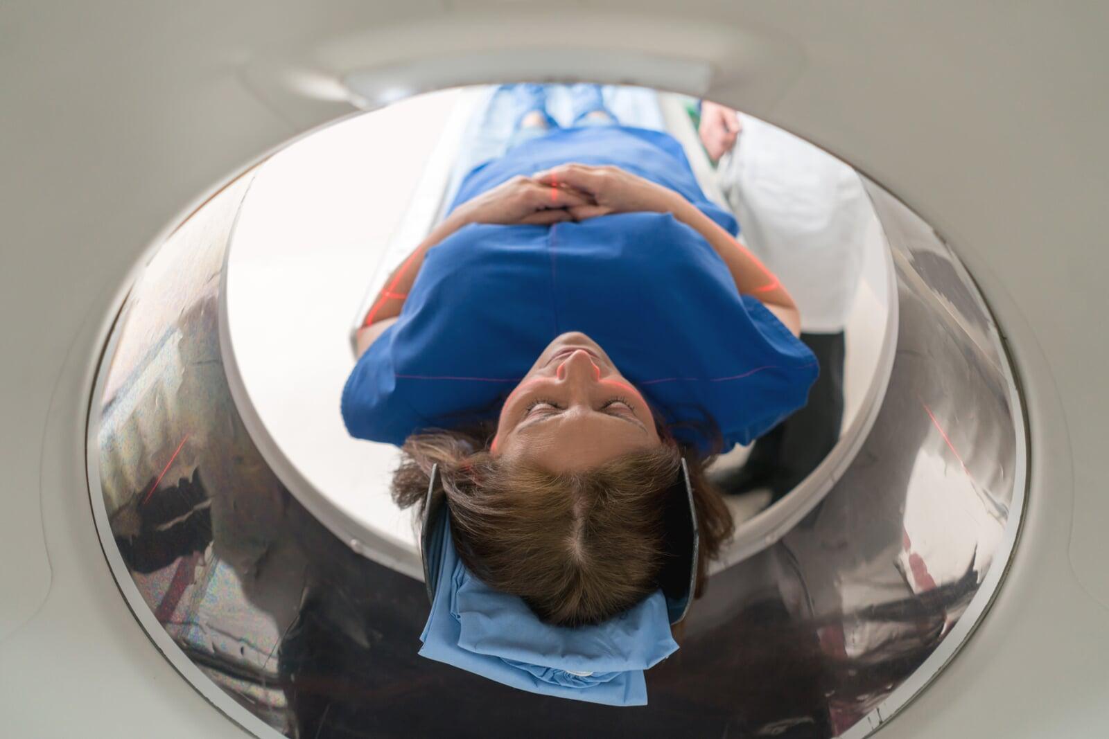
Imaging is a cornerstone of most oncology clinical trials, playing a critical role in tumor response assessment, disease monitoring and, ultimately, in evaluating therapies. However, discrepancies in imaging acquisition and interpretation across multi-center trial sites can introduce noise into the data, making it difficult to determine actual treatment effects if not acknowledged and controlled where possible.
Key sources of imaging variability in multi-center trials include:
Differences in Equipment and Settings: Variability in MRI, CT, or PET scan machines, along with differences in resolution, contrast agents, and imaging protocols, can lead to inconsistencies in tumor measurement and classification.
Variability in Image Acquisition: Differences in slice thickness, field-of-view, and scanning sequences can alter tumor appearance, leading to potential misclassification of progression or response.
Inter-Observer Variability: Differences in how radiologists interpret and measure tumor response can lead to inconsistencies in Response Evaluation Criteria in Solid Tumors (RECIST) or other criteria-based assessments.
Data Processing Differences: Image reconstruction, post-processing software, and AI-driven analytics may vary across sites, introducing additional discrepancies in tumor evaluation.
"These inconsistencies can significantly affect trial results, inaccurately assigning treatment response rates and producing misleading efficacy data or, in some cases, regulatory and approval delays that hinder patient access to promising therapies."
Strategies to Standardize Imaging Across Trial Sites
1. Workflow Technology and Software Platforms
Advances in software-as-a-service technologies establish platforms that span departments and disciplines, clinical and research divisions, across sites and various institutions to harmonize workflow. Purposefully designed, these products allow scaled collaboration and automate processes to standardize imaging assessments and reporting with ease. Built into their design are barriers to undesired human variation that would affect trial data accuracy.
2. Centralized Imaging Review and Quality Control
3. Standardized Imaging Protocols
Establishing and enforcing trial-wide imaging protocols—including uniform contrast agent use, slice thickness, and scan timing—can reduce inconsistencies across different sites, but some leniency may be necessary for institutional guidelines unless specific rationales can be given. Protocol training and regular compliance audits help ensure adherence to standardized processes once established.
4. AI and Automated Image Analysis
Leveraging artificial intelligence (AI) and machine learning tools can enhance consistency in image interpretation. AI-driven analysis can detect subtle changes in tumors more accurately, reducing human error and observer variability. This is being used more frequently, but this technology, too, needs to be appreciated for how different algorithms from different platforms may allow divergence across trial sites.
5. Cross-Site Calibration and Equipment Harmonization
Harmonizing imaging equipment settings across trial sites can reduce technical variability where possible. Regular calibration of scanners and phantom imaging studies can help maintain consistency in imaging outputs.
6. Site Training and Standardization Workshops
Conducting mandatory training sessions for radiologists and imaging technicians involved in the trial ensures uniform interpretation of tumor response criteria. Continuous education on evolving imaging technologies and standardized methodologies is essential.
"Ensuring imaging consistency across multi-center trials will enhance data reliability and accelerate drug development and regulatory approvals."
The Future of Imaging Standardization in Oncology Trials
As oncology research moves toward more personalized and targeted therapies, the need for precise and reproducible imaging data has never been greater. Ensuring imaging consistency across multi-center trials will enhance data reliability and accelerate drug development and regulatory approvals. By adopting reliable standardization methods and automating workflow, leveraging AI, and promoting centralized imaging review, clinical research professionals can play a pivotal role in driving oncology innovation forward.
The success of future cancer treatments depends on the accuracy of today's clinical trials. Reducing imaging variability across multi-center studies is not just an operational goal—it is a commitment to scientific integrity and, ultimately, to the patients who rely on these advancements for better outcomes.